Posts

Gram staining

Image
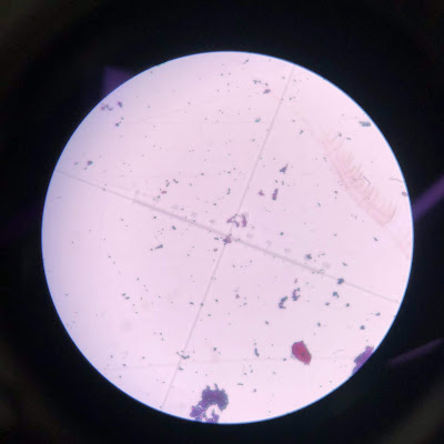
Happy sunny morning!!! I came the busy lab in Saturday morning and did few gram staining experiments for caeni 2 and caeni 3. Both of them are  Gram positive bacteria. Gram positive bacteria: Dark purple Caeni 3     Gram positive bacteria  :               Dark purple Caeni 2 

Blog for Lab's work on 3/3/2020

Image
Besides being used for stock cultures, lab uses many of these media which are excellent for experiments. TGY broth and TGY agar are excellent medi; almost students and mentors in my lab get by using it alone. This week I did the list of 500 ml TSB, 250 mL TGY broth and 250 mL TGY agar (spread to 11 dishes).. 1. Did 500 ml TSB 15 g Bacto Tryptic Soy Broth Soybean - Casein digest medium  500 mL of DI water 2.Did 250 ml TGY borth 1.25 g Tryptone 0.25 g Glucose 1.25 g Yeast 250 mL of DI water 3.Did 250 ml TGY agar 1.25 g Tryptone 0.25 g Glucose 1.25 g Yeast 3.75 g Agar 250 mL of DI water

Running the gel electrophoresis 2/21/2020 and 2/22/2020

Image
That was a successful day at lab. Our group did the good job with cutie gel and nice results.  I joined to do the  gel electrophoresis 1.8 % and ran machine at 85 mA/Rm. I had few pictures for that days:

Working with rich media and gel electrophoresis 2/14/2020 and 2/15/2020

Image
1. I did the rich media 500 mL agar with the component: + Bacto peptone 5g + Yeast extract 2.5g + Casamino acid 2.5 g + Meat extract 1g beef + Mail extract 2.5 g + Glycerol 1g + MgSO4 x 7H20 0.5g + Tween 80 0.005g + Agar 10g + DI water 500 mL 2. I also did the rich media 500 mL with the component: + Bacto peptone 5g + Yeast extract 2.5g + Casamino acid 2.5 g + Meat extract 1g beef + Mail extract 2.5 g + Glycerol 1g + MgSO4 x 7H20 0.5g + Tween 80 0.005g + Agar 10g 3. I also jointed to make gel electrophoresis 1.8%

Gram staining from 2/7 to 2/8/2020

Image
This week I did the gram staining for 13 sample of gobi and geo group in 2 days from 2/7/2020 and 2/8/2020. I did washing slides over 5 seconds so it might effect to my results. I have thought I did bad in this week for Gram staining. I will try the best in next time.

Fluorescent gram staining protocol v1

Image
Slides were loaded into a BioTek slide in both a coverslip down Optimal exposure settings were determined for each objective using a positive control smear and then applied to all images for both positive and negative controls. Our group used microscope BioTek to do gram staining protocol with sample was CF594 (red color/cell wall) and Dapi (Blue color/ Nuclei) With + DAPI 377/477 1225100 Rev J - Blue + GFP 469/526 1225101 Rev K- green + TEXAS 586/647 125102 Rev J - Red I very like this new technique, and I think it is more exactly than normal gram staining technique I have used before. I hope I will do it few more time in future so I can use to do this new method.

DNA Transformation

Image
I did the Transformation of E. cloni 10G Chemically Competent cells: 1. Remove Recovery Medium from the freezer and bring to room temperature.  2. Remove E. cloni 10G cells from the -80°C freezer and thaw completely on wet ice (10-15 minutes).  Tube (+) Tube (D) Tube (I) 3. Thaw the tube of pRham™ Vector DNA and microcentrifuge the tube briefly to collect the solution in the bottom of the tube.  4. Add 2 µL (25 ng) of the pRham Vector DNA and 1 to 3 µL (25 to 100 ng) of insert product to the cells. Stir briefly with pipet tip; do not pipet up and down to mix, which can introduce air bubbles and warm the cells.  5. Important: Transfer the mixture of cells and DNA to a pre-chilled disposable polypropylene 15- mL culture tube (17 x 100 mm). Performing the heat shock in the small tube in which the cells are provided will significantly reduce the transformation efficiency.  Summary:  Put 2 µL Prham ™ C-His + 3 µL transformation to tub...